However, research is revealing that it is far more complex than just a switchboard between your ears.The layers vary in thickness at different sites on the cortex for example, the granular layers (layers 2 and 4) are more prominent in the primary sensory area and less so in the primary motor area. The classical idea of the human brain is the command center of your nervous system. Knowledge of the clinically-oriented anatomy of the meninges is essential for all physicians and. Disruption of these functions can cause reversible and irreversible damage to the brain parenchyma. There are three distinct meningeal layers, the dura, arachnoid and pia mater, that serve key structural and homeostatic functions for the brain.
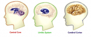
Neuroscientists have attempted to break down the different brain parts to explain its structure.He called it the triune brain and suggested that three distinct brains emerged successively in the course of evolution and now coexist in the human skull. Your brain is so powerful and so diverse, it’s a big task to name all its different functions. Dura mater: is a strong, thick membrane that closely lines the inside of the skull its two layers, the periosteal and meningeal dura, are fused and separate. From the outermost layer inward they are: the dura mater, arachnoid mater, and pia mater.

As a data-storage system, the long-term hard drive of the brain may be infinite. That equates to between 18 and 640 trillion signals that your brain sends and receives every second.On top of all this, it stores some of the information it transmits as memories. Each synapse can convey a signal at 0.1–2 times per second.
It allows researchers to explore broader data on brain scan patterns by comparing neural markers and functional outcomes.Firstly, the human brain is divided into left and right hemispheres. However, the absolute contribution of each area to behavior is yet to be properly defined.New large studies are integrating brain imaging and behavioral data. These are areas, networks, and systems.It is mostly known that the brain is organized into functional areas that are integrated into networks. It’s organized at many physical scales starting at the level of single neurons and expanding up to functional systems.Current functional imaging studies classify brain structure to three levels. It is believed that the brain can remember everything from birth through to death.The cerebral cortex is what sets apart human brains from other organisms.
One hemisphere may be slightly dominant. Then the right brain controls the left side. The two sides are strongly though not entirely symmetrical.The left brain controls muscles on the right-hand side of the body.

Some researchers think this area is subdivided into two or three sections. V2 sends feedback connections to V1 and has feedforward connections with V3-V5.V3 is located immediately in front of V2. Cells in this region respond to color, spatial frequency, moderately complex patterns, and orientation. This is called the occipital lobe and is located in the left and right hemispheres.It integrates and processes visual data relayed from the retinas.It’s divided into five different areas (V1 to V6) based on function and structure.V1 respond to specific types of visual cues such as the orientation of edges and lines.V2 receives and processes data from V1. It sets us most apart from other creatures.The primary visual cortex is found at the back of the human brain. It’s also known as the cerebral cortex and controls many of the thoughts and processes we equate to the human mind.
Unlike V2, V4 is tuned certain details of complexity, like simple shapes.V5 thought to play a major role in the perception of motion, the integration of local motion signals into global percepts, and the guidance of some eye movements.V6 appears to respond to visual stimuli associated with self-motion [and wide-field stimulationPart of the temporal lobe of the cerebral cortex. Like V2, V4 is tuned for orientation, spatial frequency, and color. Ventral V3 (VP), has primary visual area links, and stronger connections with the inferior temporal cortex.V4 is a lesser understood area involved with selective attention.
Supplementary motor cortex – controls movements requiring two muscle groups. Primary motor cortex – sends signals the motor neurons of the spinal cord to stimulate the intended muscles. Eg, to kick a soccer ball, it the right leg muscle group must be used. Premotor cortex – chooses the muscle group required for an action. The left motor cortex controls the right side of the body, and right motor cortex controls the left side of the body. The motor cortex sends muscle signals down the spinal cord.


 0 kommentar(er)
